
Adapting imaging instruments to shed new light on plant biology
September 17, 2021
Austrian BioImaging/CMI
Plant Biology
User stories
Imaging living plants as they grow has traditionally been very difficult as almost all microscopes place the sample horizontally but plants grow vertically. This challenge has been overcome by scientists at Austria’s Institute of Science and Technology, part of Austrian BioImaging, CMI, Euro-BioImaging’s Austrian Node. With a “special” confocal microscope for vertical sample mounting and integrated directional illumination, combined with a custom software for tracking moving objects, live plant imaging becomes easy. This technology, and related expertise, are available to all scientists, regardless of affiliation, via the Euro-BioImaging portal. Learn more in this interview with Gabriel Krens, Manager of the Bioimaging Facility at Austria’s Institute of Science and Technology.
Your institute has developed a microscope specifically for imaging plant roots as they grow. Please tell us a bit more about the capabilities of this system.
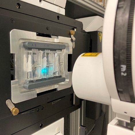
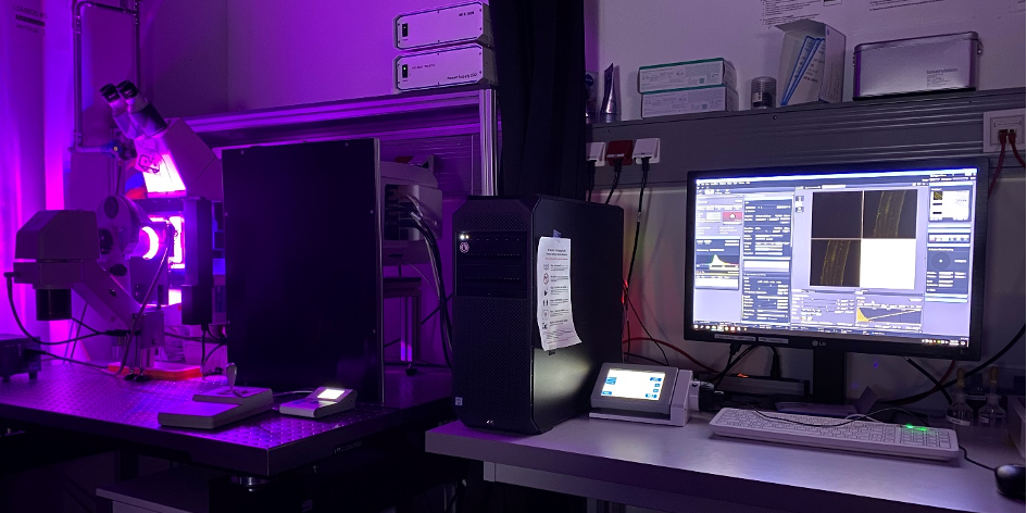
Gabriel Krens: The goal of the researchers from the Benkova and Friml research groups is to follow molecular players in the process of root and stalk growth over time on a subcellular level. As plant development is steered by gravity, the samples – Arabidopsis thaliana – cannot be mounted in a horizontal manner to study the process of interest. To overcome this problem, a first version of the vertically mounted confocal microscope has been established in the facility by Ekaterina Papusheva and Robert Hauschild, based on a LSM700 system. This version made use of custom adaptor plates to position the microscope, as well as in-house developed scripts that allowed to stabilize the acquired images of the growing root over time while recording its displacement. By doing so, the growth speed can be tracked, while the subcellular processes can be recorded with confocal resolution over time. Over the years, the system has been upgraded and now has different holders that hold to up to 3 root-chips that each can house over 20 roots; additional microfluidics and rotational stages have been developed; light stimulation accessories have been added; the scripts have been updated, and a second system with sub-diffraction limited imaging resolution modality have been added.
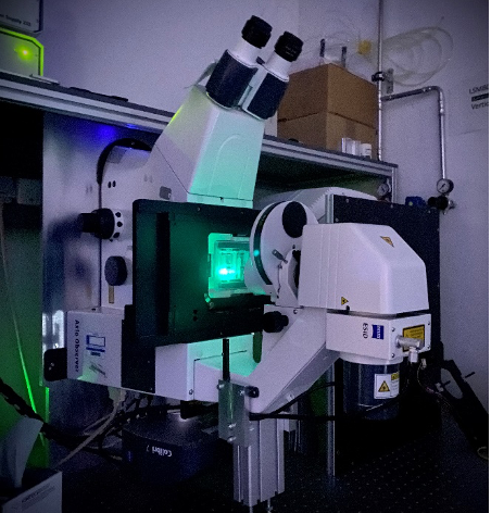
Specification Vertical confocal 1, Zeiss LSM-05 (800 vertical), at IST Austria can be found here.
Specification vertical confocal 2, Zeiss LSM-06 (800 vertical + Airyscan), at IST Austria can be found here.
Are there any particular challenges with this new microscope orientation? What things do you have to pay special attention to?
Gabriel Krens: We have an excellent mechanical workshop that helps us with developing the required adapters, as well as the additional sample holders, rotational stage and lights. One challenge that was unknown beforehand was if we could correct the scanner orientation after placing the microscope in the vertical position. In addition, the later upgrade of the microscope also came with challenges, as we needed to reestablish the online analysis scripts. This certainly came with significant effort and required patience to overcome those issues. Nevertheless, all the new implementation allow for easier adaptations for further automation, whereby we are able to respond for possible alterations that are required for new experiments.
What kind of projects do you support using this technology? What scientific questions are you be able to address?
Gabriel Krens: As we work in a facility, we aim to provide a service for the scientist. On this specific setup, we are mostly providing services and custom solutions to researchers working on plant development. The rotational stage makes it possible to abruptly change the direction of gravitational forces. The roots respond to such changes by adapting the direction of growth along the newly imposed gravitational axis. Our vertical microscope(s) allows us to follow subcellular processes involved in this behavior. In addition, researchers are able to cancel-out gravitational forces by systematically rotating the sample. These application are of course not limited to plant samples. More recently, the system has been equipped with a micro-fluidics setup that allows to flush-in different treatments. The effects of such treatments can then again be monitored by confocal imaging.
What other services do you provide in your facility that would be useful in combination with this type of microscopy?
Gabriel Krens: The vertical confocal microscopes have the same functionality as the similar inverted confocal microscopes and thus all applications that can normally be performed – or applications that can be combined on any inverted confocal microscope - can also be done on the vertical microscope, as long as the sample is compatible with the vertical mounting. To increase the compatibility for additional samples, we developed several custom adapters to secure samples’ safety.
An exciting example of a combination of services used on the vertical confocal microscopes was done in a follow-up study, where laser-assisted cell ablation were combined with confocal imaging on the vertical microscopes. Several exciting findings have recently been published where the imaging of wound-healing responses in plant roots was followed upon laser ablation, as well as new insights into regulation of growth by the plant hormone auxin have been unraveled.
Why should scientists choose your facility for using this technology?
Gabriel Krens: Generally, we facilitate the maintenance of state-of-the-art microscopy and flow cytometry equipment, advanced user training, assay development, image analysis and provide project-based solutions for optical development. Specifically, when a scientific project requires an imaging application that would require a vertically mounted sample, or if there is an interest to study the effect of gravity, the vertical confocal microscopes are an established tool that provide unique insights.
About Austrian BioImaging
The Austrian BioImaging Node CMI (Correlated Multimodality Imaging Node) is a multi-sited, multimodality Node covering biological and biomedical imaging from cryo-electron and advanced light microscopy up to the preclinical level (including microCT, microMRI, microPET). It is hosted by 8 leading institutions across Austria with a broad service offer for organic materials, biomedical model organisms and humans, numerous multimodality imaging pipelines, and various support services, such as data and image analysis. Imaging technologies at Austrian BioImaging/CMI can be combined or used as stand-alone technologies depending on the specific biomedical research question of the user. Learn more here.
Learn more about live plant imaging:
Read the complete peer-reviewed article: https://elifesciences.org/articles/26792#media1
References to follow-up studies involving wound-healing studies:
Hoermayer L, Montesinos JC, Marhava P, Benková E, Yoshida S, Friml J. Proc Natl Acad Sci U S A. 2020 Jun 30;117(26):15322-15331. doi: 10.1073/pnas.2003346117. Epub 2020 Jun 15.
Re-activation of Stem Cell Pathways for Pattern Restoration in Plant Wound Healing.
Marhava P, Hoermayer L, Yoshida S, Marhavý P, Benková E, Friml J. Cell. 2019 May 2;177(4):957-969.e13. doi: 10.1016/j.cell.2019.04.015.
More news from Euro-BioImaging

March 12, 2025
EVOLVE Job Shadowing Stories: Paula Jiménez Gómez visits the DIMP NEUROMED - leadership, measuring impact and the challenges of project coordination.
The EVOLVE Job Shadowing program is a fantastic opportunity for Node staff to immerse in the daily life and operations of other Nodes of…