
September 13, 2024
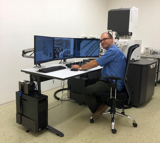
Innovative CryoEM Workflow Development: An In-Depth Look at TESCAN and the Institute of Molecular Genetics Collaboration
At the Special Edition Virtual Pub, Dominik Pinkas (Institute of Molecular Genetics of the Czech Academy of Sciences, part of Euro-BioImaging’s Advanced LIght & Electron Microscopy Prague Node) & Ondřej Šulák (Tescan), will present a use case on how they worked together in an academia-industry partnership to develop and optimise workflows for cryo-electron microscopy. Their talk is entitled “Innovative CryoEM Workflow Development: An In-Depth Look at TESCAN and the Institute of Molecular Genetics Collaboration.”

February 21, 2024
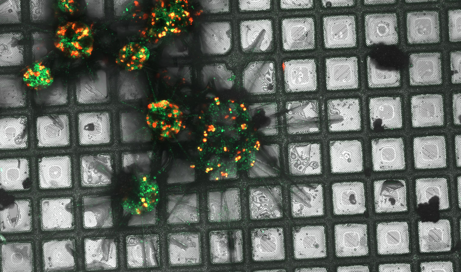
Dissecting the natural history of the Malaria parasite Plasmodium falciparum in mosquitoes with advanced 3D electron microscopy
Pablo Suárez Cortés is a Postdoctoral researcher at the Max Planck Institute for Infection Biology, Berlin (Germany). His work focuses on understanding how Plasmodium falciparum,…


February 13, 2023
Volume EM Series: 3D Correlative Live and Cryogenic imaging of Biological Tissues combining Raman, Light and Electron Microscopy
On Friday, February 17th at 13:00 CET, Nico Sommerdijk, Department of Medical Biosciences, Electron Microscopy Center, Radboudumc Technology Center Microscopy, Radboudumc, Nijmegen-NL delivers a…
