2024 will stand out in France-BioImaging history as a very positive year. Not only was France-BioImaging awarded significant national funding to support their infrastructure…
June 17, 2024
New facilities join Euro-BioImaging Nodes
We are delighted to welcome new facilities into the Euro-BioImaging family as part of our recent Node upgrade. Four Euro-BioImaging Nodes – Swedish NMI,…
March 11, 2024
Providing customised bioimage analysis advice & workflows through a nationwide, remotely operating core-facility
Many Euro-BioImaging Nodes are working on innovative image analysis and image data management solutions and the next Euro-BioImaging User Forum is designed to highlight…
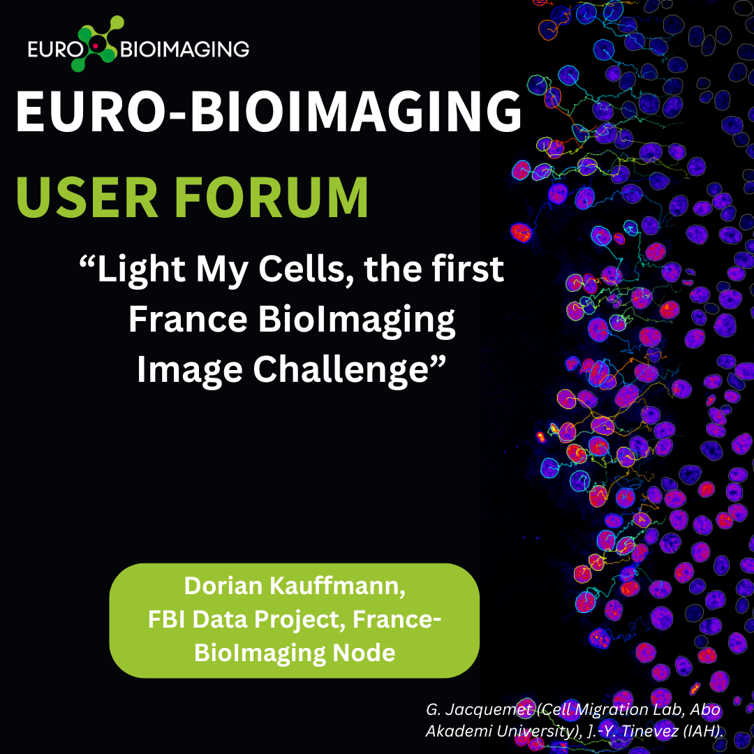
March 11, 2024
Light My Cells, the first France BioImaging Image Challenge
Join us at the Euro-BioImaging User Forum, for an afternoon of presentations from Euro-BioImaging users and image data/image analysis experts at our Nodes that…
January 26, 2024
Successful AI Basics for Image Analysis webinar available on YouTube
In November-December 2023, the Horizon Europe-funded ANERIS Project organized a workshop series to explore the realms of AI application for image processing. These workshops,…
October 25, 2023
Super-resolution imaging inside brain slices with lattice light-sheet microscopy
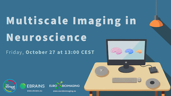
Our next Special Edition Virtual Pub, “Multiscale Imaging in the Neurosciences,” organized in collaboration with EBRAINS, will take place on Friday, October 27. At…
September 7, 2023
Correlative Light and Electron Microscopy of Plant tissues
Imaging technologies support research into the structure and function of plants, shed light on plant health, resilience and adaptability, and help answer agroecology-related research…
March 23, 2023
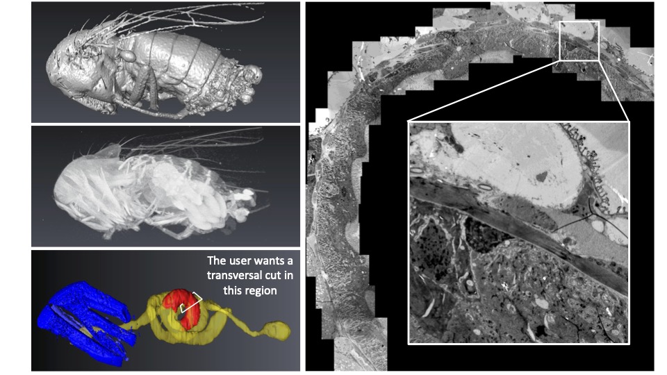
CXEM: Finding a needle in a haystack
Correlative X-ray imaging and electron microscopy (CXEM) is the combination of X-ray imaging and electron microscopy. It is a correlative approach that makes it…
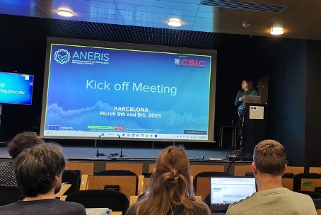
March 8, 2023
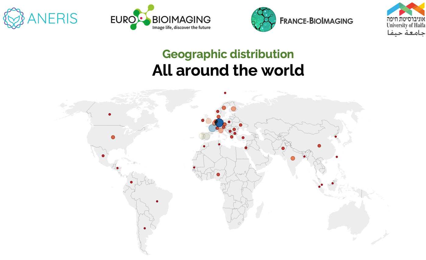
ANERIS Project: Towards Operational Marine Biology
Euro-BioImaging is delighted to be part of the ANERIS (operAtional seNsing lifE technologies for maRIne ecosystemS) project, which kicks off this week in Barcelona. The…
March 7, 2023
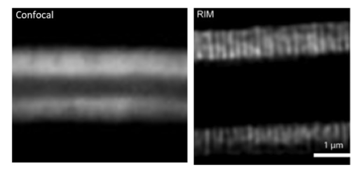
A powerful high speed, low phototoxicity microscopy method to achieve super-resolved images
Are you interested in looking at tissues or other thick samples in high resolution? We spoke to Marc Tramier, a group leader at the…
December 12, 2022
Image data analysis services at France BioImaging
Today, imaging scientists are producing datasets of increasing volume, complexity, and information content. To realize the full potential of imaging data, Euro-BioImaging Nodes offer…
November 15, 2022
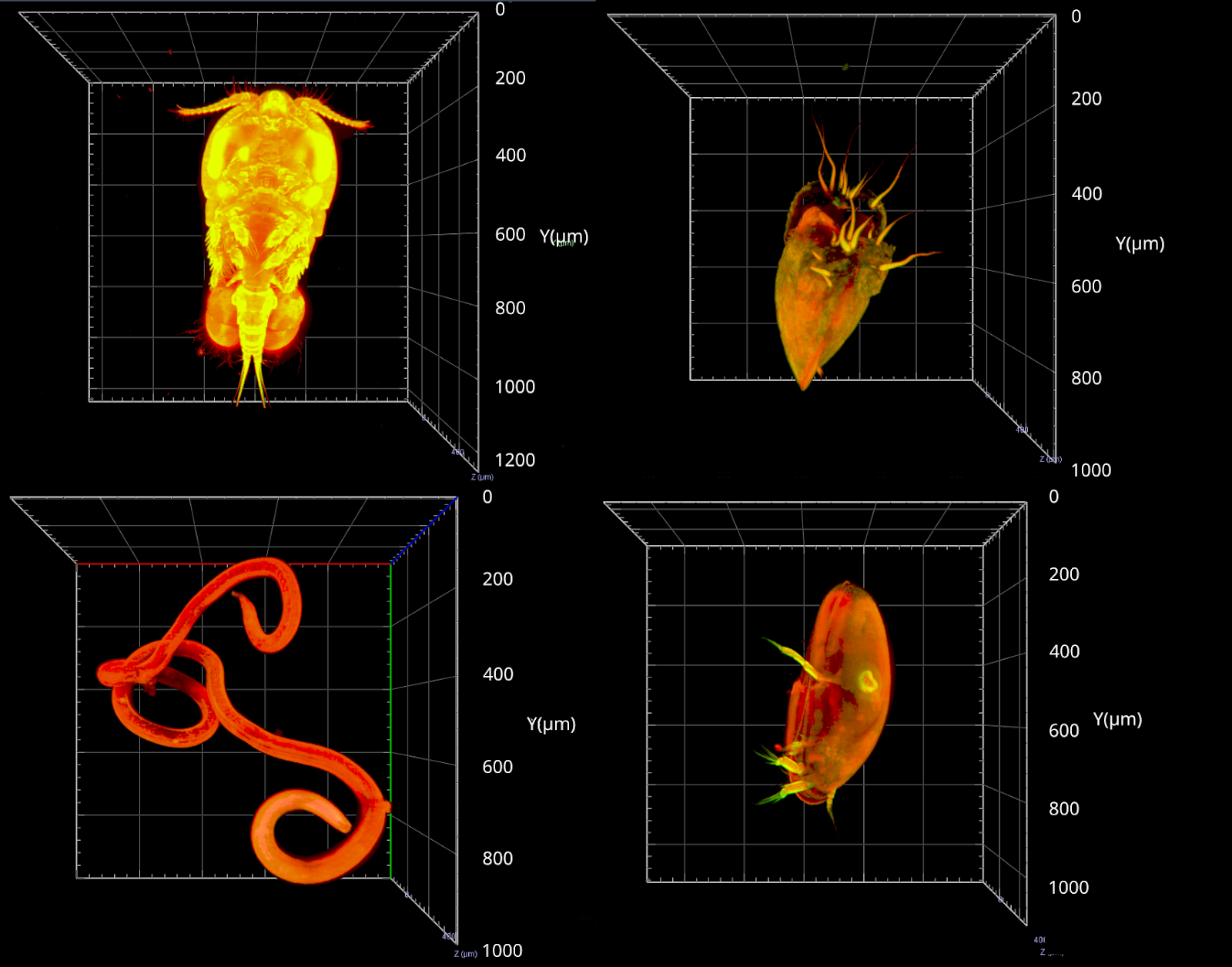
Meiofauna – the ocean’s next frontier
Meiofauna are tiny marine organisms that range in size from 20 microns to 1 milimetre. They are present everywhere in the sea, from the…