Fantastic ELMI 2023 Conference in Noordwijkerhout!
This year, the European Light Microscopy Initiative meeting took place in Noodwijkerhout, the Netherlands, organised by NL-BioImaging. Several Hub team members were able to…
June 15, 2023
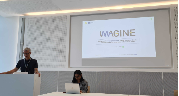
IMAGINE project – imaging technology developments to address socio-economic challenges
Euro-BioImaging is pleased to be part of the IMAGINE project, which kicked off this week at the EMBL Imaging Centre in Heidelberg, Germany. The…
June 7, 2023
Supporting the development of hydrogels for drug delivery
Francois Lux is an Associate Professor of Chemistry at the University of Lyon 1. His research interests include nanomedicines – precisely the development of…
May 23, 2023
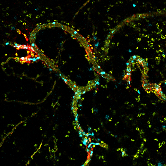
Intravital microscopy supports progress in nanomedicine and immunotherapy
Understanding how immune cells interact with different medications within the disease environment is at the heart of Alexandros Marios Sofias research interest. He is…
May 23, 2023
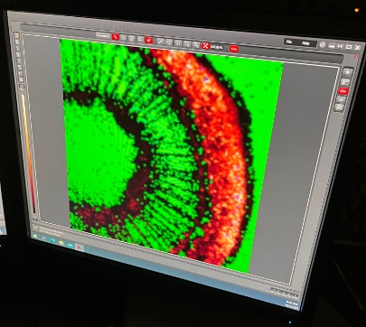
Looking at stem photosynthesis with FLIM
It all started at the Molecular and Biophysical Bases of Photosynthesis Conference held in Venice in May 2022. Sara Natale, then a PhD researcher…
May 19, 2023
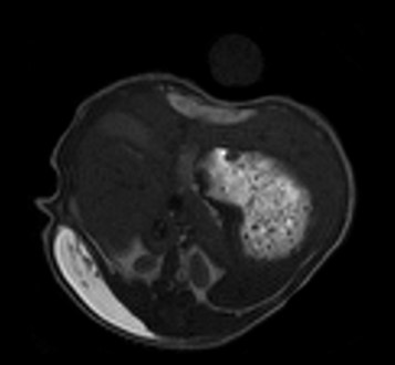
Understanding Phantom Limb Pain
Phantom Limb Pain (PLP), i.e., the pain perceived in an amputated body part, is a very debilitating condition for amputated patients. However, its origin…
May 5, 2023
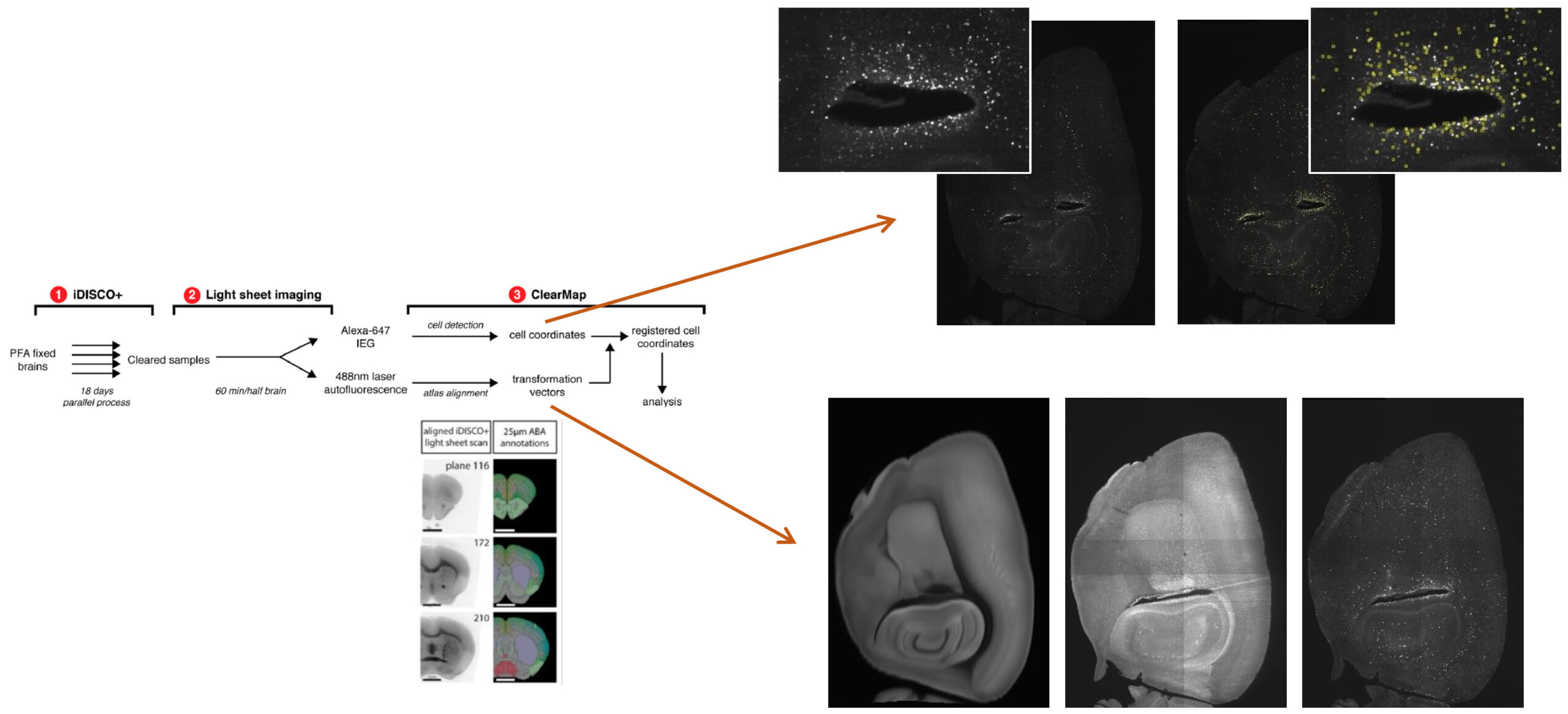
Analyze large-scale light-sheet microscopy images of the brain
Want to analyze and annotate your sample with the Allen Brain Atlas but don’t have the hardware, software or know-how…
April 5, 2023
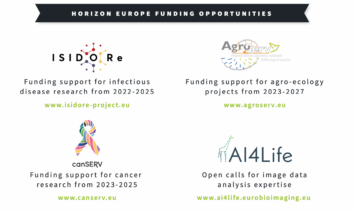
Get funding to visit Euro-BioImaging imaging facilities
Euro-BioImaging ERIC is a European research infrastructure consortium (ERIC) that offers open access to state-of-the-art imaging technologies, training, and data services in biological and…
April 3, 2023
Spotlight on Austria – March 2023
From March 13-17, 2023, several Euro-BioImaging team members had the opportunity to travel to Austria for EMIM 2023. On their way to Salzburg, they…
March 28, 2023
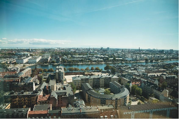
WORLD OF IMAGING - Euro-BioImaging visits the Danish BioImaging Node
Euro-BioImaging is delighted to share some stories from our fantastic Nodes. First on the long list is the Danish BioImaging Node (DBI). Euro-BioImaging visited…
March 23, 2023
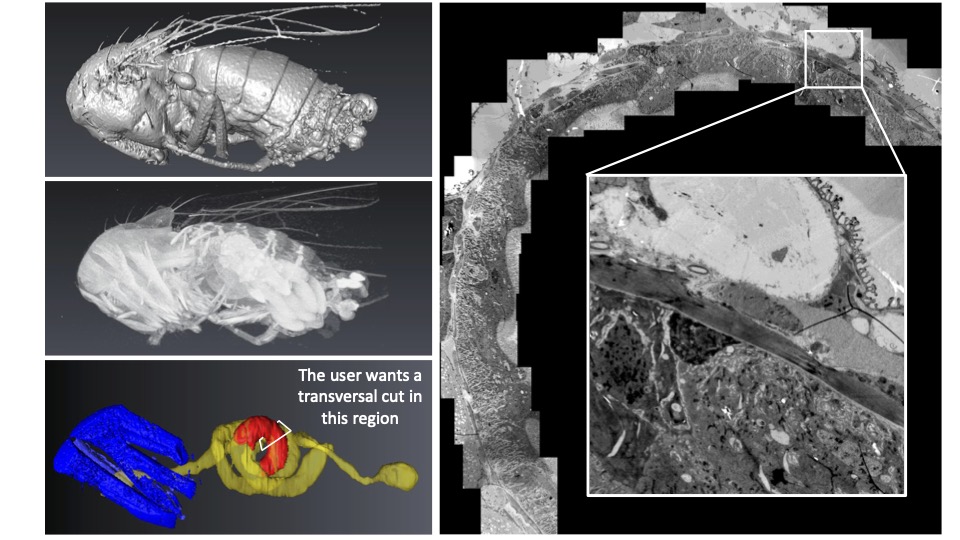
CXEM: Finding a needle in a haystack
Correlative X-ray imaging and electron microscopy (CXEM) is the combination of X-ray imaging and electron microscopy. It is a correlative approach that makes it…
March 16, 2023
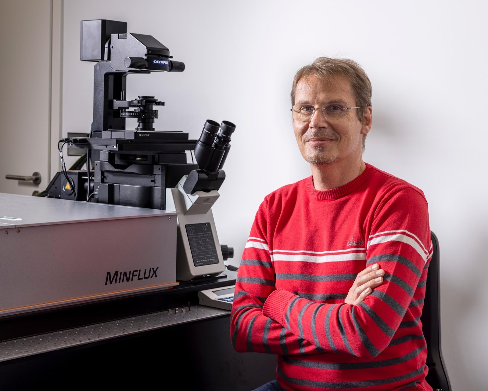
MINFLUX: A light microscopy technique that closes the gap on structural biology
MINFLUX is a super-resolution approach developed by Nobel prize laureate Stefan Hell in 2016. Two Euro-BioImaging Nodes are currently offering MINFLUX in open access…