EDX image at 4 nm pixelsize showing phosphor, osmium and sulfur distribution in EPON-embedded exocrine pancreas, in red, green and blue respectively. Image credit: Peter Duinkerken, Giepmans lab, UMC Groningen, NL.

January 17, 2024

EDX Imaging: Electron Microscopy beyond the grey-scale

Electron Microscopy holds the key to understanding the ultrastructure of biological systems, but finding what you are looking for in the high resolution EM…

Maximum intensity projection of Acantharia (plankton), plunge frozen and imaged with a high-end confocal microscope. Green: Acantharia; red: chlorophyll of symbionts (microalgae). Credits: Anna Steyer/EMBL

January 3, 2024

Cryo-ET: Imagination is the limit

Studying macromolecular complexes in their native environment in a cell is a real challenge, but new methods and technology advances are allowing scientists to…

The lightweight, transportable TeraHertz Imaging system offered by the University of Pisa can easily be deployed in the field.

November 20, 2023

TeraHertz Imaging: Portable plant imaging

Working on plant biology? Want to detect leaf water stress or assess the quality of dry fruits like chestnuts and hazelnuts? TeraHertz Imaging could…

Like a ball of yarn… Above is an example of how CXEM can be used to find the best itinerary within a sample to make transverse sections of the portion of interest. On the left, the micro-CT provided a 3D map of the sample within the resin block. On the right, a transverse ultrathin TEM section of the drosphilia gut. Image courtesy of Nuno Luis (Schnorrer lab, IBDM) & Nicolas Brouilly (Electron Microscopy Facility, IBDM AMU/CNRS, France BioImaging).

March 23, 2023

CXEM: Finding a needle in a haystack

Correlative X-ray imaging and electron microscopy (CXEM) is the combination of X-ray imaging and electron microscopy. It is a correlative approach that makes it…

Timo Zimmerman, team leader for light microscopy services at the EMBL Imaging Center. Photo courtesy of EMBL PhotoLab.

March 16, 2023

MINFLUX: A light microscopy technique that closes the gap on structural biology

MINFLUX is a super-resolution approach developed by Nobel prize laureate Stefan Hell in 2016. Two Euro-BioImaging Nodes are currently offering MINFLUX in open access…

Dr Selene Roberts at the MINFLUX machine at the OCTOPUS facility, part of Euro-BioImaging's UK Node.

March 14, 2023

MINFLUX: Super fast, super-resolution microscopy

Want to see objects that are 5 nanometers apart? Need to track objects moving in live cells with high temporal resolution? Then why not…

Laser Microdissection at the Polish Advanced Light Microscopy Node.

March 9, 2023

Laser Microdissection: Extract specific regions from your sample

Want to look at tumor vs. healthy tissue within a sample? Laser microdissection enables you to select a material based on its spatial or…

RIM comparison

March 7, 2023

A powerful high speed, low phototoxicity microscopy method to achieve super-resolved images

Are you interested in looking at tissues or other thick samples in high resolution? We spoke to Marc Tramier, a group leader at the…

Spatial trasncriptomics. Image courtesy of Bioscience Technology Facility, University of York.

March 1, 2023

Spatial Transcriptomics – For understanding tissue architecture

Spatial Transcriptomics is a useful approach for understanding tissue architecture and for understanding the molecular basis of health and disease. The Bioscience Technology Facility…

Magenta: membrane coated beads, green: virus like particles. Image courtesy of Steven Edwards, SciLifeLab.

February 28, 2023

Lattice Light-sheet: For fast, subcellular, volumetric imaging

Want to see adherent cells with low phototoxicity and photobleaching? What about spheroids and organoids? Both of these are possible with Lattice Light-sheet imaging,…

Example of Expansion Microscopy. Image courtesy of Ana Agostinho, SciLifeLab, Swedish NMI.

February 23, 2023

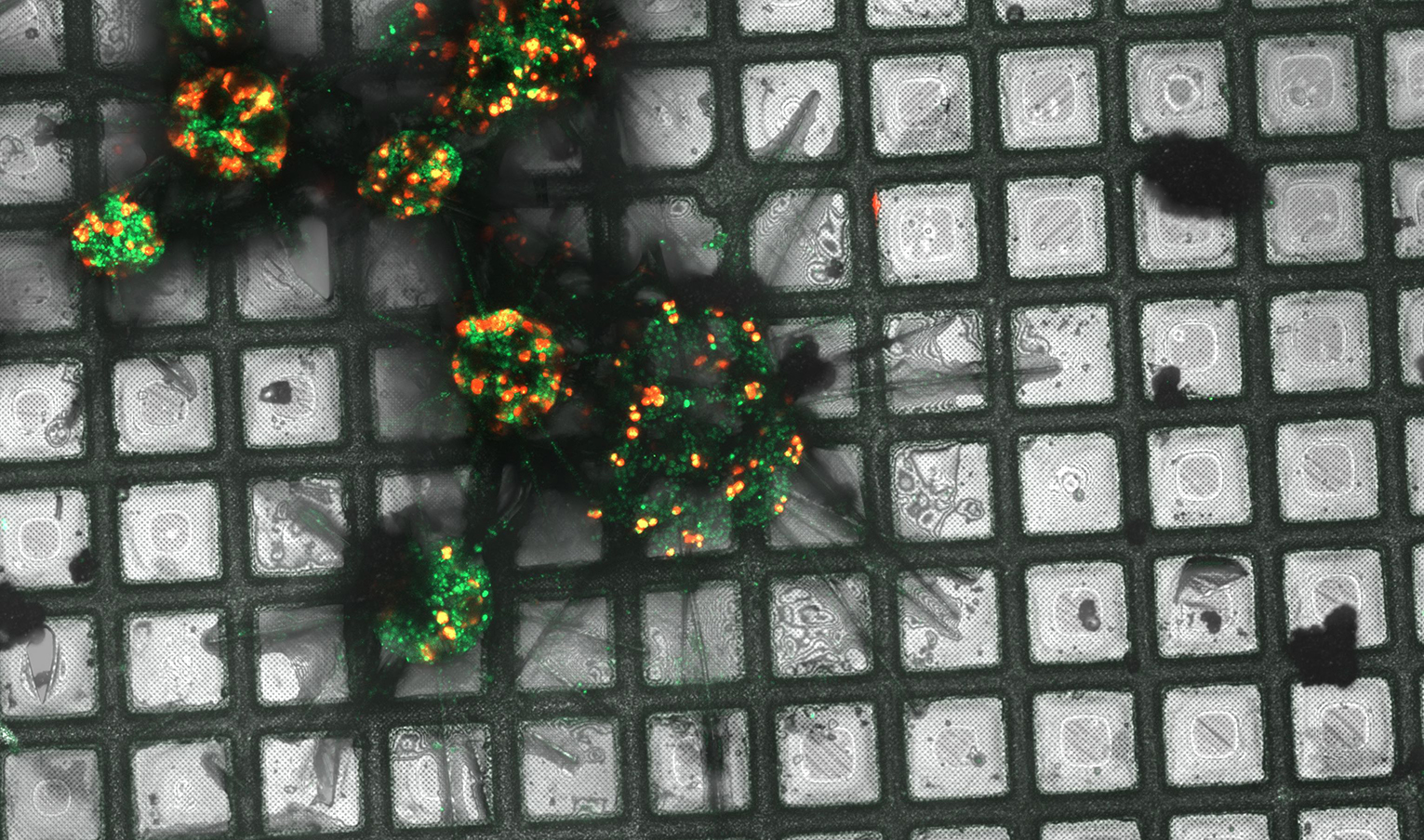
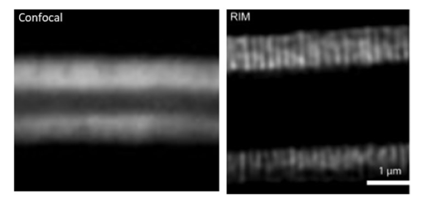
Expansion Microscopy: Enabling super-resolution microscopy to be performed on diffraction limited microscopes

Want to look at subcellular structures such as mitochondria, centrioles and microtubules in super-resolution? Only have access to conventional diffraction-limited microscopes? By physically expanding…