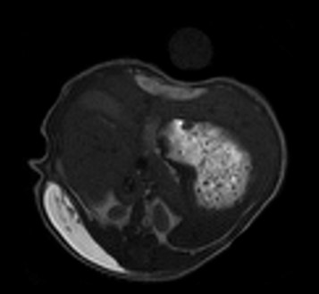
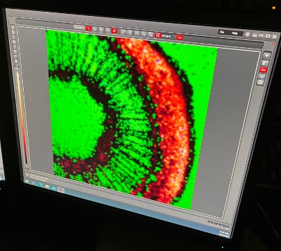
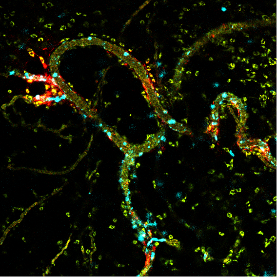
Multiscale multimodal 3D analysis of cardiovascular alterations/ structural features in a rhesus macaque monkey model for COVID-19
The ongoing COVID-19 pandemic is the biggest health crisis of the 21th century. Due to the extremely high number of infections, it is of…
June 16, 2023
Interested in Smart Microscopy? Join the discussion!
Towards community standards in Adaptive Feedback Microscopy Adaptive Feedback or ‘Smart’ Microscopy is an emerging field that is gaining importance e.g. in the context…
June 15, 2023
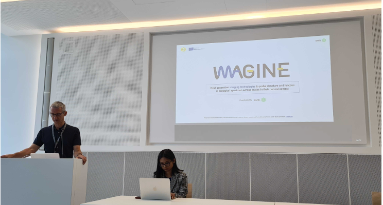
IMAGINE project – imaging technology developments to address socio-economic challenges
Euro-BioImaging is pleased to be part of the IMAGINE project, which kicked off this week at the EMBL Imaging Centre in Heidelberg, Germany. The…
June 15, 2023
Join our Special Edition Virtual Pub: Showcasing Stories & Research from ISIDORe Users
On Friday, June 23, at 13:00 CEST we are organizing a Special Edition Virtual Pub with our ISIDORe users. The session will highlight the…
June 12, 2023
Funding for Correlated Multimodal Imaging projects from COMULISglobe
COMULISglobe, a CZI-funded project, aims at consolidating and extending a collaborative and innovative network that promotes MultiModal Imaging and analysis across scales (MMI) from…
June 8, 2023
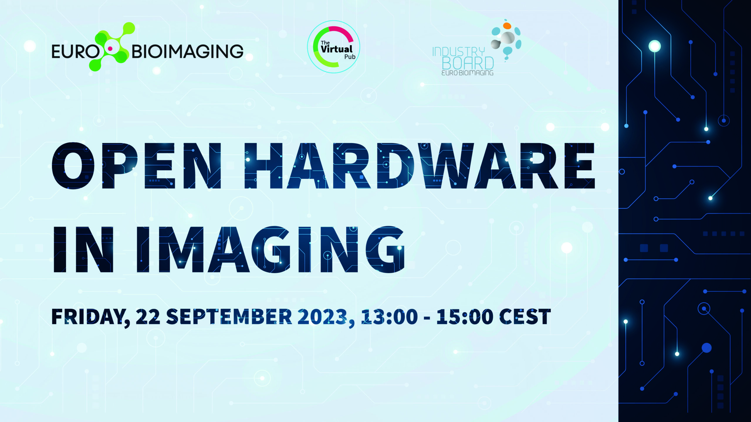
Submit your Abstract: Open Hardware in Imaging
Imaging technologies are becoming increasingly complex and ever more expensive, reducing the general accessibility and potential reach of cutting-edge techniques. Many scientists and companies…
June 7, 2023
Supporting the development of hydrogels for drug delivery
Francois Lux is an Associate Professor of Chemistry at the University of Lyon 1. His research interests include nanomedicines – precisely the development of…
June 6, 2023
Euro-BioImaging is at ELMI 2023!
This week, Euro-BioImaging Hub team members will attend ELMI 2023 in Noordwijkerhout, The Netherlands. We are planning lots of opportunities to meet up during…
May 23, 2023
Looking at stem photosynthesis with FLIM
It all started at the Molecular and Biophysical Bases of Photosynthesis Conference held in Venice in May 2022. Sara Natale, then a PhD researcher…
May 23, 2023
Intravital microscopy supports progress in nanomedicine and immunotherapy
Understanding how immune cells interact with different medications within the disease environment is at the heart of Alexandros Marios Sofias research interest. He is…
May 19, 2023
Understanding Phantom Limb Pain
Phantom Limb Pain (PLP), i.e., the pain perceived in an amputated body part, is a very debilitating condition for amputated patients. However, its origin…
May 16, 2023
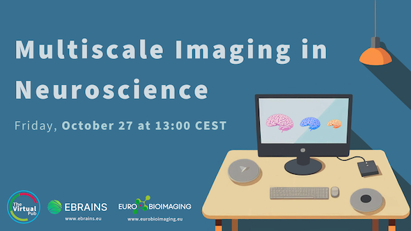
EBRAINS – Euro-BioImaging workshop series
Euro-BioImaging ERIC and EBRAINS are Research Infrastructures that offer complementary services to neuroscientists, namely advanced imaging technologies for neuroscience, and…