Touring Austrian BioImaging – Or Scientific Sightseeing in my Hometown
October 14, 2024
Austrian BioImaging/CMI
Nodes
Scientific Ambassadors
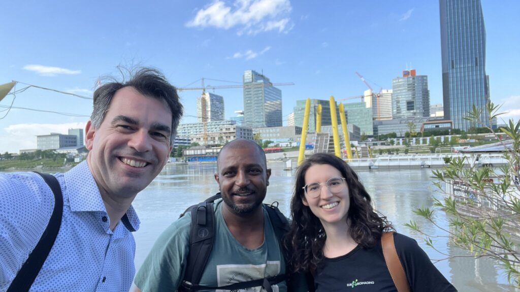
In June 2024, Euro-BioImaging Scientific Ambassador, Magdalena Schindler, a native of Vienna, had the opportunity to visit Austrian BioImaging/CMI, Euro-BioImaging's Austrian Node, in a whirlwind tour organised by Baubak Bajoghli, Operating Director of Austrian BioImaging/CMI. She shares some highlights from her seven stops in a five-day tour in the article below. More insights from her tour are published on the Austrian BioImaging/CMI website.

Magdalena Schindler
Magdalena Schindler is a Predoctoral Fellow at the European Molecular Biology Laboratory (EMBL) in Heidelberg. Her research focus is on mechanobiology in developmental processes. Her enthusiasm for microscopy and collaborative research led her to apply to be one of Euro-BioImaging’s first Scientific Ambassadors in 2024. As part of her role, Magdalena, known as Lena, connected with Euro-BioImaging colleagues in her hometown of Vienna. Some insights from her amazing experience in Vienna are published below.
Visiting just one-third of the Austrian Node in five days left a lasting impression of the openness and passion shared by its members. It also highlighted how important their association with Euro-BioImaging is to them.
- Magdalena Schindler, predoctoral researcher, EMBL
Article written by Magdalena Schindler
As a Euro-BioImaging Scientific Ambassador, I recently had the opportunity to explore Austrian BioImaging/CMI, meeting various members of the Austrian Node. Baubak Bajoghli, Operating Director of Austrian BioImaging/CMI, kindly arranged a tour of several associated imaging core facilities and research groups. I was very impressed by the array of cutting-edge imaging technologies and the dedicated staff supporting researchers. During my tour, I got the chance to talk to several members working as heads, senior scientists, or facility managers in core facilities or research groups. Links with details of their work are provided within the text. Here, I share my experiences and highlights from my seven stops in a five-day tour.

Stop 1 - Vienna Biocenter Core Facilities (VBCF)
My trip started at the VBCF (1 on the map) where the Austrian Node’s head office is currently located. Its director Baubak explained to me the structure of Austrian BioImaging/CMI, a consortium of eight universities and research institutes, including 29 imaging core facilities and research groups that offer access to 35 biological and 14 preclinical imaging technologies and bioinformatic services.
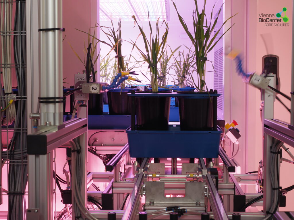
At VBCF, I then visited two core facilities that are part of Austrian BioImaging/CMI: the PHENOplant and the Electron Microscopy Core. At PHENOplant, Jakub Jez showcased their advanced infrastructure for high-throughput, fully automated plant phenotyping under various environmental simulations and controlled stresses. The facility welcomes external users, with services potentially funded by the AgroServ project. At the Electron Microscopy Core, I observed sample preparation and data acquisition for scanning and transmission electron microscopy.
Stop 2 - University of Vienna
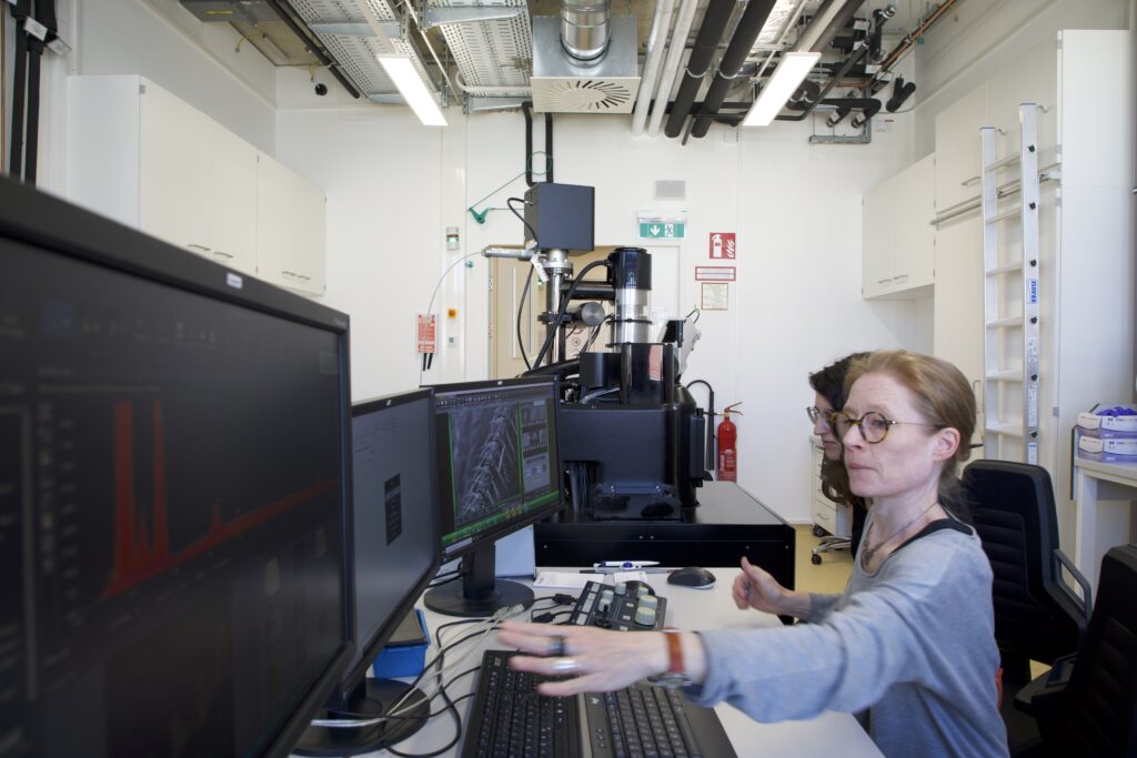
Next, I crossed the road to the new University of Vienna Biology Building (UBB) to visit Katy Schmidt and her microscopy core facility (2). UBB is equipped with several light and electron microscopy instruments, specializing in imaging microorganisms, plants, and insects with high resolution. Katy showed us some fantastic electron microscope images of a butterfly and an ant analysed down to their atoms through elemental dot mapping. After this lesson on insect physiology, Katy shared her passion for supporting scientists over a nice cup of coffee with us. Throughout her career, he has worked with researchers from various backgrounds and affiliations.
Stop 3 - TU Wien (formerly: Vienna University of Technology)
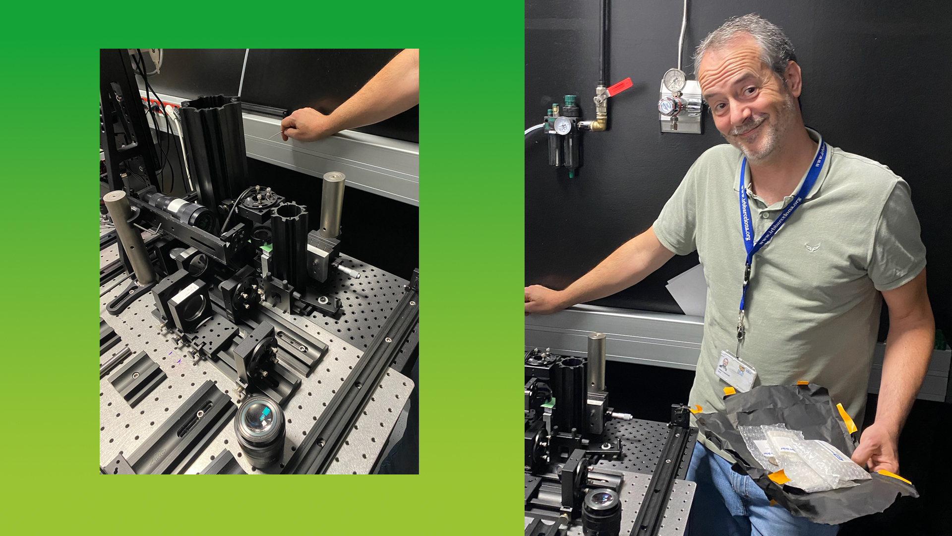
On the second day, I visited 3 research groups associated with the Austrian Node, all located in Vienna’s inner city. A few tram stops from the Vienna State Opera, I was welcomed by Victor Weiss at the TU Wien (3). He is part of the Research Group for Mass Spectrometric Bio and Polymer Analytics led by Martina Marchetti-Deschmann. Martina’s team provides access to Mass Spectrometry Imaging (MALDI-Imaging) through Austrian BioImaging/CMI. I enjoyed hearing about the origin story of their imaging technologies, starting with samples from chicken legs purchased at the “Naschmarkt”, the oldest market in Vienna with over 200 years of history, located just 100 meters away from the institute. The group has since developed numerous multimodal mass spectrometric imaging methods also facilitating multi-instrument approaches, collaborating with both academic and industry partners like Shimadzu. MALDI imaging and services that Martina’s team offer to the external users are part of the canSERV and ISIDORe service catalogues, two European projects that provide researchers financial support to access cutting-edge research infrastructures and services.
Right next door, I visited Orestis Andriotis, a senior scientist in the research group Biomechanics led by Philipp Thurner at TU Wien. We discussed their extensive nano- and micromechanics techniques including atomic force microscopy (AFM), with experiments ranging from nanoscale collagen fibrils to cell clusters and tissues. They also have a strong expertise in combining AFM with other imaging modalities, such as Stochastic Optical Reconstruction Microscopy (STORM), MALDI imaging or Brillouin Scattering microscopy (BSM) in collaboration with other groups within the Austrian Node (read also my interview with Orestis). The two groups I met at TU Wien have combined their techniques to reveal both chemical composition and mechanical properties of samples by correlating AFM with MALDI imaging.
Stop 4 - Medical University of Vienna
After a brief lunch, I visited the Department of Anatomy and Cell Biology of the Medical University of Vienna not far off Vienna’s prestigious ring road (4). Here, I first met with Kareem Elsayad, whose group has developed new techniques in Brillouin Scattering Microscopy (BSM) to characterize the physical properties of biological materials (read also my interview with Kareem). You can find some of his recent work in PNAS and Nature Photonics.
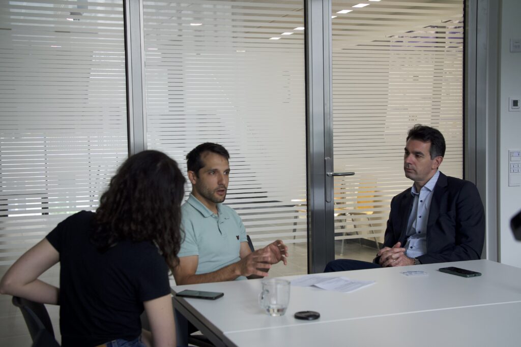
Next, I got the opportunity to interview Wolfgang Weninger, the head of the Anatomy Department and the Scientific Director of the Austrian BioImaging/CMI. After I had heard quite a bit about collaborations from individual scientists, Wolfgang put this in a wider context. He explained how these close collaborations within the Node’s research groups and imaging core facilities have led to the development of various protocols and methods to combine up to seven image modalities from a given sample. That is why CMI as an expertise of the Node is embedded in its name.
Stop 5 - VRVis
On the third day, I ventured across the Danube River to Donau City, a modern part of Vienna, and home to the VRVis Research Centre for Virtual Reality and Visualisation (5). At VRVis I met Katja Bühler, Scientific Director of VRVis and Head of the Biomedical Image Informatics Group. VRVis specializes in visual computing solutions for industry, biological research, and medical applications, with a focus on machine learning-powered image analysis tools. Thus, they are building a bridge between academic research, industry and medicine. Katja is keen to get her hands on any kind of data, as long as there is an interesting problem to solve. She told me about project data ranging from microscopy and medical imaging to satellite images of crop fields. As machine learning can be used for many of these problems, understanding its reliability and reproducibility is essential. Katja wants to ensure reliability and quality standards that are necessary for applications of this, currently, black box in clinical as well as research and industry contexts. I really enjoyed this real-world application focused development of machine learning in image analysis, since up to then I had seen this technology mostly at more preliminary stages in research talks.
Stop 6 - Vienna Veterinary University
My last stop in Vienna was the University of Veterinary Medicine Vienna (6), the third oldest veterinary institution in the world. I met Ursula Reichart and Stephan Handschuh, two senior scientists in the team of imaging specialists led by Martin Glösmann at the VetCore Facility for Research. Ursula and Stephan showed me several state-of-the-art microscopes, among them two super-resolution and one spinning disk microscope as well as X-ray micro-CT. Stephan explained to me that the core facility is fully open to external users, with access to workstations for image data analysis. Ursula told me that users are supported in experimental design and trained in the use of hardware and software, but “knowledge transfer plays a key role, and we motivate scientists to contribute to image acquisition and analysis themselves.” For some technologies, such as X-ray micro-CT, the core facility also offers a comprehensive service. In this case, users can send in their samples and the core staff takes care of the imaging. The facility also has a great deal of expertise in working with samples from different animal species. Ursula told me: “As we are based at a vet school, we process different samples, not only from mice or rats, but also from dogs, cats and horses.” “We routinely combine tomography with downstream microscopy, either light or electron microscopy,” explained Stephan. When instruments for certain workflows are not available on campus, analysis takes place at other sites in Vienna, and sample transportation between facilities is coordinated by Austrian BioImaging/CMI, which facilitates the combination of imaging modalities.

Stop 7 - ISTA
On my last day, I traveled to Klosterneuburg, a historic town just 15 km from Vienna, to visit ISTA (Institute of Science and Technology Austria; 7). I got a tour of the imaging and optics facility by image analysis expert Tereza Belinova, during which I learned about ISTA’s advanced imaging technologies and data infrastructure. Gabriel Krens, the head of the facility, emphasized the importance of creating an overarching infrastructure for image data sharing to foster more efficient and responsible research. He hopes Austrian BioImaging and Euro-BioImaging will play a key role in this effort. This lovely sentiment concluded my trip.
Conclusions
Visiting just one-third of the Austrian Node in 5 days left a lasting impression of the openness and passion shared by its members. It also highlighted how important their association with Euro-BioImaging is to them.
I want to thank Euro-BioImaging for the wonderful opportunity to serve as its ambassador; Baubak Bajoghli for organizing the visits; Marianna Poli for her support; Henok Karvonen for taking professional photos and all members of the Austrian Node who took the time to meet with me and share their passion for imaging.
More news from Euro-BioImaging