Technologies
Overview
Through the large number of included facilities, Euro-BioImaging can offer access to the full range of imaging technologies in the biological and biomedical imaging field. Our technology portfolio covers everything from the nano- to the tissue- and organism scale. We are constantly adding new technologies - making sure that the latest cutting-edge imaging technologies, such as MINFLUX and spatial transcriptomics, are available in open access to all researchers.
You can browse our technologies here.
Harnessing the imaging revolution
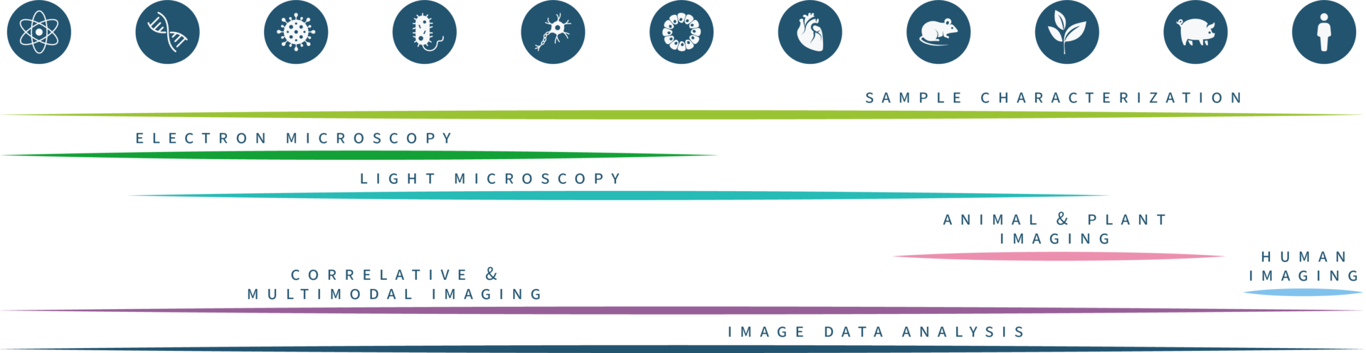
The Euro-BioImaging technology portfolio ranges from light and electron microscopy on the biological imaging side to an expanding range of applications of biomedical imaging, from plant and ex-vivo imaging to animal and human imaging applications.
Electron Microscopy
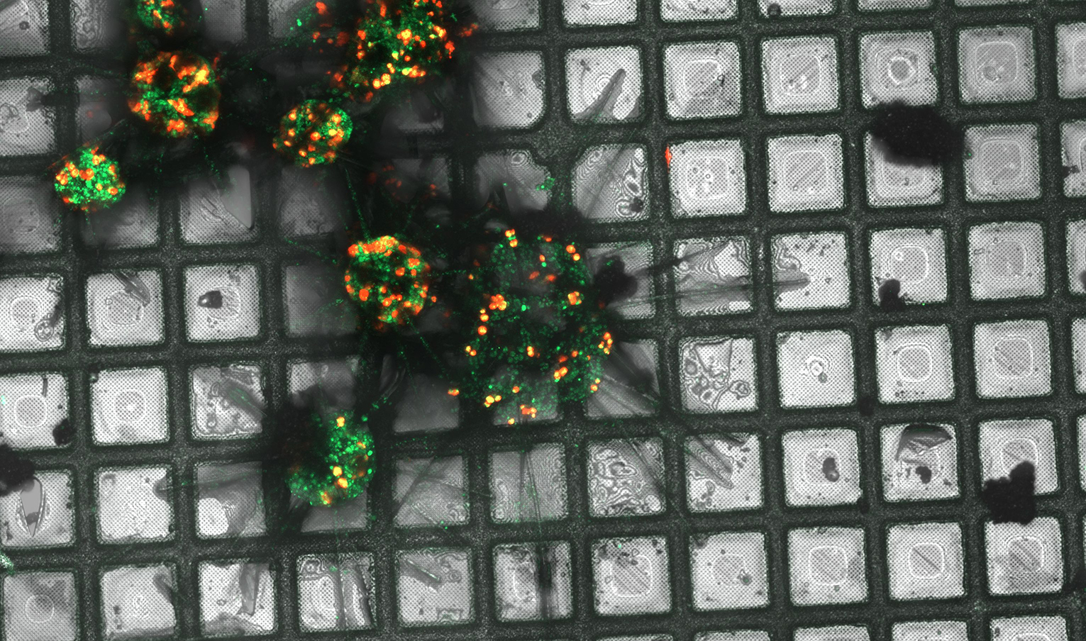
Our Electron Microscopy portfolio covers cryo-EM techniques for in situ structural exploration, such as cryo-electron tomography (cryo-ET), as well as the full complement of volume EM techniques, such as FIB-SEM, Array Tomography and Serial Blockface SEM. Many of our facilities also specialise in correlative methods, Correlative X-ray Imaging and EM (CXEM) and Correlative Light and Electron Microscopy (CLEM).
Left: Maximum intensity projection of Acantharia (plankton), plunge frozen and imaged with a high-end confocal microscope as part of a CLEM workflow. Green: Acantharia; red: chlorophyll of symbionts (microalgae). Credits: Anna Steyer/EMBL
Light Microscopy
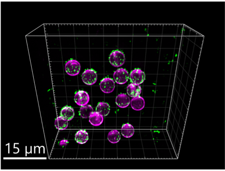
In light microscopy, our Nodes offer everything from confocal microscopy up to single molecule location approaches and intravital imaging. Our light microscopy techniques allow for 3D live cell imaging, tracking, high content screening, and include a variety of functional imaging techniques to explore protein dynamics in living cells. Recently we have added a number of new and highly requested methods, such as MINFLUX, Single Particle tracking, Spatial transcriptomics, and Lattice Lightsheet microscopy to our portfolio.
Right: Imaged with Lattice Lightsheet, in magenta membrane coated beads and in green virus like particles. Image courtesy of Steven Edwards, SciLifeLab.
Multimodal in vivo animal and human imaging

Left: 68Ga-DOTA-Siglec-9 PET/CT image of hands of a 49-y-old patient with early Rheumatoid Arthritis (Finnish Biomedical Imaging Node)
Our preclinical imaging Nodes provide access to all the relevant biomedical imaging technologies, from Magnetic Resonance Imaging to Ultrasound, Optical and PhotoAcoustic Imaging, up to the nuclear imaging technologies (PET, SPECT, CT and bimodal techniques) for animal studies with applications in preclinical research (e.g. evaluation of new therapeutics, assessment of new molecular probes and imaging markers) and in veterinary science. Expertise on protocols, sequences, probes and tracers is also provided where needed. Some of our Nodes also provide access to technologies for studies on human subjects. In this case support for subjects recruitment and ethics management is also provided.
Model Systems
Euro-BioImaging facilities also offer access and support with imaging a wide range of model systems and can advise how to get the best imaging results out of them, from Drosophila, zebrafish, mouse embryos and organoid systems to small rodents and even bigger animal models of the various pathologies. Here access to instruments is complemented by technical expertise of facility staff to support specialized sample and animal handling.
Adaptive technologies to explore all facets of your sample
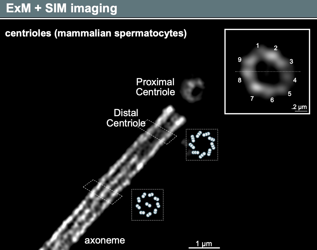
And of course, we also provide access to adaptive and support technologies, such as laser-based microdissection, Feedback Microscopy, high-speed imaging, microscopes at high biosafety levels, and specialized sample preparation methods, such as Tissue Clearing and Expansion Microscopy.
Euro-BioImaging can also support you if you want to explore the physical and chemical properties of your samples - through access to a range of methods such as MassSpec Imaging, Atomic Force Microscopy, and chemical imaging, such as µ-XRF and μ-PIXE.
Left: Example of a centriol imaged with Structured Illumination Microscopy (SIM) after Expansion Microscopy sample preparation. Image courtesy of Ana Agostinho, SciLifeLab, Swedish NMI.